�������ǰǧ�����ʳ�������ڲ�����ռ����Ҫϯλ�������о����������������t�W�����I��Ҳ���F�������������к����S���Ĵ��͵V���|Ԫ�أ��Ƃ��^���в������κ��ж�ԇ�������ﰲȫ�����á����ڶ�����ˇ�_�l�Ĵ��M������֧�ܾ߂䶹�����õ����ﰲȫ�ԣ�ͬ�r�߂����ɶ�ăȲ��Y����һ�������W���ܣ�����ͬ�r�����˴����W�������Ƃ�Ĵ��M������֧�ܵ����|���������M����ϵ�y���о����״Ό�����֧���������t�W�I���Б����M���˳���̽���������_�l�������������t�W�I��ĝ��ڃrֵ��
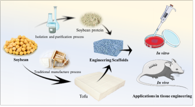
Figure 1. Schematic illustration of developing tofu scaffolds and soybean protein scaffolds
���գ���ɽ��W���WԺ�����t�W���̌W�ƅ��x�n�}�M�״Ό����y�Ķ����_�l�ɽM������֧�ܣ��������������t�W�I���еđ����M���˳���̽�����@�nj�ʳԴ����������_�l����һ�δ�đ�Lԇ�������Ԃ��y�����Ƃ乤ˇ����A���ڲ������ж�ԇ����ǰ���������Ƃ�ɿɑ����ڽM�����̵�����֧�ܣ�������ܛ�M�������ޏ��аl�]һ�����á����ɹ���Evaluation of tofu as potential tissue engineering scaffold ���}�����x�������־�i���о��T��ͨӍ���ߣ��S�E��ʿ���һ���ߣ���ɽ��W���WԺ���һ��λ����2018��1��23�հl���ڲ��όW�����ڿ���Journal of Materials Chemistry B��(DOI: 10.1039/C7TB02852K) �ϡ�
ԓ�Fꠌ����y�����Ƃ乤ˇ�M�к��θ��������ڶ���֧�ܵ��_�l����Ч�������ж�ԇ�������룬�O��������˲��ϵ����ﰲȫ�ԣ�ͬ�r�������˴����W�������Ƃ�Ĵ��M������֧�ܵ����|���о�����������֧���c���W�������Ƃ�Ĵ���֧�ܾ������Ƶ��������ܣ��Һ��ڣ�����֧�ܱ��F�����ѵ����������ԣ��������ڼ�������ֳ���w��ֲ���Ѫ�ܵ��γɡ�
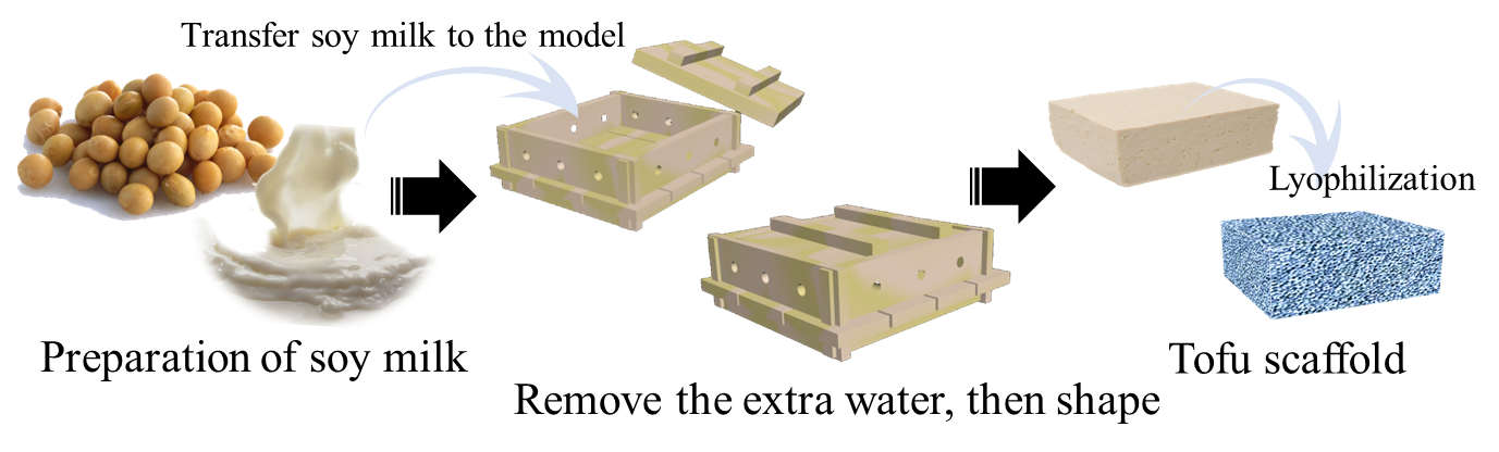
Figure 2. The simplified process of tofu scaffold preparation.
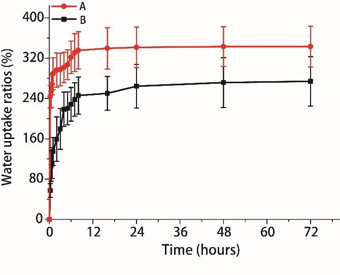
Figure 3. The water uptake ratios of tofu scaffolds and soybean protein scaffolds in 0.01 M PBS solution: (A) Tofu scaffolds. (B) Soybean protein scaffolds. Mean �� SD��n=3.
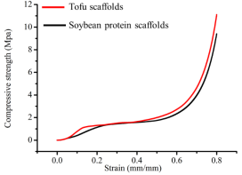
Figure 4. The stress�Cstrain curves of tofu scaffolds and soybean protein scaffolds under wet conditions. There was no significant difference to the compression modulus of tofu scaffolds and soybean protein scaffolds. Mean �� SD, n = 3
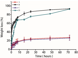
Figure 5. The result of weight-loss ratio of soybean protein and tofu scaffold: (A) Tofu scaffold in 0.01 M PBS solution. (B) Tofu scaffold in 0.25% trypsin solution. (C) Soybean protein scaffold in 0.01 M PBS solution. (D) Soybean protein scaffold in 0.25% trypsin solution. Mean �� SD, n = 3.
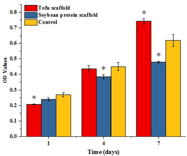
Figure 6. Proliferation of NIH 3T3 cells after 7 days culture with the leaching solution of soybean protein and tofu scaffolds. ��p < 0.05 (compared with the control at the same time), Mean �� SD, n = 3.

Figure 7. The morphology of 3T3 cells directly seeding in soybean protein and tofu scaffolds were analyzed by staining with rhodamine phalloidin and DAPI (A, B, C, D). Live/dead staining was carried out to verify the good biocompatibility of two scaffolds (E, F, G, H). (A&E): The morphology of 3T3 cells seeded in soybean protein scaffolds after cultivating 24 hours. (B&F): The morphology of 3T3 cells seeded in soybean protein scaffolds after cultivating 48 hours. (C&G): The morphology of 3T3 cells seeded in tofu scaffolds after cultivating 24 hours. (D&H): The morphology of 3T3 cells seeded in tofu scaffolds after cultivating 48 hours. The cell morphology is good after seeding in two types scaffolds directly, which were indicated by blue arrows. The dead cells are indicated by white arrows. Scales bar = 50 ��m.

Figure 8. The results of histology evaluation implanting tofu scaffolds (A, B and C) and soybean protein scaffolds (D, E and F) after 7 days. (A & D): HE stained section of the scaffold with surrounding tissues. Tofu scaffolds part was indicated by blue arrows and good biological integration with the surrounding tissues of implantation site was observed, which represented good biocompatibility of tofu scaffolds; (B & E): Masson staining displayed obvious collagen deposition in tofu scaffold part, indicated by yellow arrows; (C & F): CD31 staining was carried out to evaluate the neovascularization. The brown spots, indicated by black arrows, represented the angiogenesis in tofu scaffolds. Many minute vessels had grown out. Scale bar = 100 ��m.
���������Ԃ��y�����Ƃ乤ˇ����A���������_�l�ɽM������֧�ܡ��о�����������֧���c���W������֧�ܾ������Ƶ��������ܣ��������̈́������������֧�ܾ������õ����������ԣ��m�ϼ��������L��ֲ���w�Ⱥ�СѪ�ܵ����ɡ�
Փ��朽ӣ�
Evaluation of tofu as potential tissue engineering scaffold��J. Mater. Chem. B, 2018, DOI: 10.1039/C7TB02852K.��
http://pubs.rsc.org/en/content/articlelanding/2018/tb/c7tb02852k#!divAbstract
�n�}�M��B��
�n�}�Mؓ؟�˅��x��������ɽ��W���WԺ�����t�W���̌W�Ʋ�ʿ��������2015���нM������ǧ��Ӌ���@���ߡ����Ͼ���W�������~�s����ʯϪ��W�@�ÌWʿ���Tʿ�Wλ��2010���������Π���W�����t�W����ϵ�@��ʿ�Wλ��2010-2015�������������W�t�WԺ����ʡ�����WԺOmid Farokhzad ��Robert Langer����ҏ��²�ʿ���о����о��I���漰�ɽ���������ϣ�ˎ��ݔ�ͣ��M�����̼��{���t�W��ǰ���������ں���������ϵ������t�W���á��n�}�Mؓ؟���Ѱl��SCIՓ�ij��^68ƪ�����е�һ����ͨӍ����28ƪ�������ôΔ����^3800�Ρ�
�n�}�M������������ϣ��{���t�W�������t�W����Ҫ�о����{���t�W������Ҫ���_�l��������ɽ��⣬���ﰲȫ�ĸ߷��ӹ��ܾۺ��������ܸ��dˎ�{�����ˎ��ݔ���wϵ�����c���[���ί�����Ѫ�ܼ������ڷ��oˎ�еđ��á������t�W������Ҫ�����������ܲ��ϣ��Y�ϼ{���g�c�ɼ������g����������֧�ܲ��ϣ����ڽM���������ޏ͡��\����־֮ʿ�����҂��������߿ɰl�����]�䣺[email protected]���n�}�M�W퓣� http://wulab.bme.sysu.edu.cn/
- �㽭��W���L�ЈF� Prog. Mater. Sci.�����ڽM���ޏ͵����m����������� 2025-11-30
- ��h�������tɏ�n�}�M Coordin. Chem. Rev.��������������� - �Ļ��Wԭ���������OӋ 2025-09-23
- ������W��Ԫ����Adv. Mater.���C�������ڽM������O��ӿڵ�������ϻ��w�S��ֲ��ʽ̽� 2025-06-27
- �п�Ժ�z���l����������/�w���� ACS Nano�����������w�S��������p�����Sͻ�����������ʻ� 2023-12-15
- �㽭��W�������n�}�M�����ӴνY�����ߵV�ﺬ��֧�ܲ����ڹ��ޏ��еđ��� 2023-05-01
- �Ϻ����������½��ڣ����ҽ��ࣩ�n�}�M�M��������Ƹ��ʿ�������� 2021-10-18